EN
Root perforation repair

Why choose FrankoLab:

  1. 01 Accurate diagnosis and treatment under a microscope
  2. 02 MTA and bioceramics — materials with proven effectiveness
  3. 03 We take on complex cases that others have turned down

Closure of a root perforation in Ivano-Frankivsk

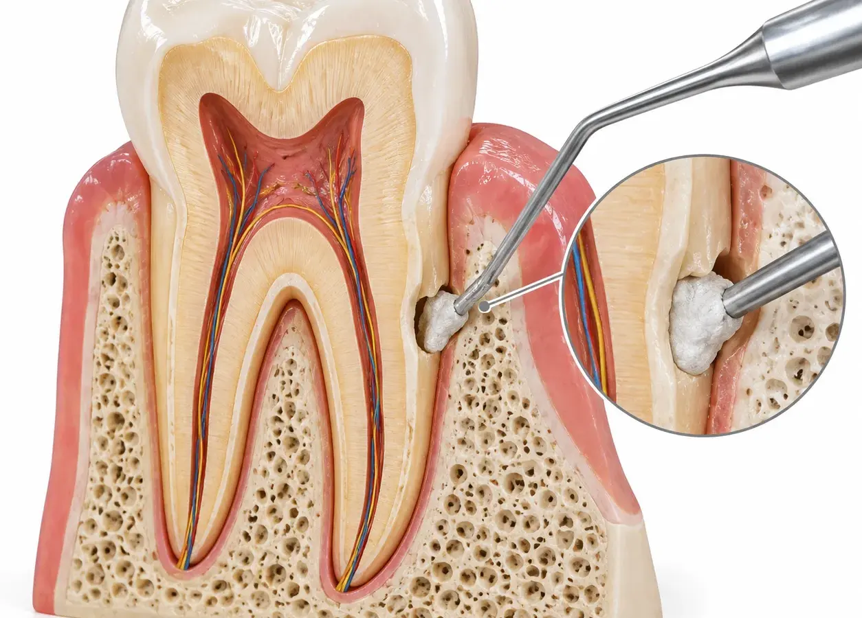
A root perforation is a pathological hole or defect in the root wall or the floor of the pulp chamber, which connects the interior of the tooth to the surrounding tissues. A perforation occurs as a result of an error during root canal treatment, trauma or the progression of caries deeper into the tooth. If not treated promptly, a root perforation becomes a gateway for infection: bacteria penetrate the bone tissue, inflammation develops, and the tooth is at risk of extraction. The earlier the defect is diagnosed and treatment begins, the greater the chances of saving the tooth.

Closing a root perforation is an endodontic procedure aimed at hermetically sealing the defect and eliminating inflammation before it leads to irreversible damage to the periradicular structures. At FrankoLab Clinic, we carry out the procedure using biocompatible materials under a Carl Zeiss / Leica / Scanner microscope.

The main causes of tooth root perforation

Errors during root canal treatment

Incorrect access to the pulp chamber, locating the root canal orifices, excessive enlargement of the canals with instruments, incorrect determination of canal length, and working in severely curved or calcified canals.

Preparation of a root canal for a post or a post-and-core restoration

One of the most common causes (according to some sources, accounting for over 50 per cent of all iatrogenic perforations) is perforation occurring during the preparation of the root canal for an intraradicular post, caused by deviation from the canal’s axis or excessive enlargement.

Root canal retreatment

Tooth root perforation occurs particularly frequently during the removal of old filling materials, posts or instrument fragments.

Lack of knowledge or complexities of dental anatomy

Failure to take into account the individual characteristics of the root’s structure, its inclination, wall thickness and the location of the canals leads to errors when creating access, locating the entry points or treating the canals, which significantly increases the risk of root perforation.

Deep caries

Gradual destruction of hard tissues (deep damage extending to the root) up to perforation of the wall or floor of the cavity.

Root resorption (internal or external)

Pathological resorption of root tissue from the inside or outside as a result of inflammatory processes, trauma or idiopathic causes.

Tooth injury

There is a possibility that a strong impact or pressure may cause cracks, fractures, root splitting or other mechanical damage, resulting in a pathological connection between the root canal and the surrounding tissues.

Additional risk factors include complex root anatomy (severe curvature, narrow canals), canal calcification, insufficient dentist experience, and carrying out fillings without proper diagnosis (X-rays, CT scans) prior to treatment.

How much does it cost to repair a perforated tooth root?

The cost of closing a root perforation at FrankoLab Clinic in Ivano-Frankivsk depends on the location and size of the defect, the chosen method of closure, the condition of the surrounding tissues, and the extent of any accompanying root canal treatment or crown restoration. We provide a transparent quote following a consultation and diagnosis, with no hidden additional charges during the process. If required, you can arrange a payment plan — please check the terms with the administrator.

The dentist will quote the final cost following an examination and diagnosis. Payment in instalments is available. Full price list for the clinic

Stages of closing a tooth root perforation

01

Diagnosis of tooth root perforation

We carry out targeted X-rays and CT scans directly at our clinic to accurately determine the location, size and nature of the defect. We assess the condition of the surrounding bone tissue and the degree of inflammation. Diagnosis under a microscope allows us to visually confirm the damage and assess the feasibility of closing a perforation in the tooth root.

02

Preparation and anaesthesia

We administer anaesthesia (local or general), use a rubber dam to isolate the tooth and ensure sterile working conditions, and, if necessary, remove old fillings or crowns to gain full access to the affected area.

03

Perforation treatment

We disinfect the perforation site and surrounding tissues. If there is active inflammation, we carry out a temporary filling using an antibacterial agent and arrange a follow-up appointment once the inflammation has subsided.

04

Closure of a perforation

Under a surgical microscope, we hermetically seal the defect with a filling material, selecting the method depending on the location and size of the perforation. We check the quality of the root perforation seal both visually and via X-ray.

05

Tooth restoration

Once the perforation in the tooth root has been sealed, we complete the root canal treatment (if required), fit a permanent filling or refer the patient for prosthetic treatment, which can be carried out at our clinic without the need to find another dentist.

06

Monitoring and dynamic observation

Once treatment is complete, we schedule follow-up appointments with X-ray diagnostics to assess tissue healing, the integrity of the perforation closure and the overall condition of the tooth. If necessary, we adjust our recommendations regarding oral hygiene and the load placed on the tooth.

Methods for closing root perforations

The choice of method for closing a root perforation depends on its location, size and how long the defect has been present.

MTA (Mineral Trioxide Aggregate)

The gold standard for closing perforations. This material, based on a mineral trioxide aggregate, has high biocompatibility, stimulates tissue regeneration and ensures a reliable seal, even in a moist environment. We use it for almost all types of root perforations.

Bioceramics (Biodentine, TotalFill BC RRM)

Modern, next-generation materials with properties similar to MTA, but with a shorter setting time. Effective for sealing perforations in both the crown and root regions of the tooth.

Surgical closure

We use this procedure for perforations in the lower third of the root or in cases where an orthograde (through-the-canal) approach is not possible. We perform the procedure as an apicoectomy or guided bone regeneration.

A combined approach

In cases of large or chronic perforations involving bone destruction, closing a root perforation sometimes requires a combination of medication, regenerative materials and surgical intervention.

Our patients’ results

Case Study 3: Composite Restorations

Diagnosis:

The patient presented with complaints of discolouration of her front teeth, numerous old composite restorations and an unsatisfactory smile. During the clinical examination, discolouration of the teeth was identified, along with restorations that had lost their functionality and aesthetic appearance.

Treatments:

Following digital planning, a decision was made to carry out aesthetic restoration of the upper incisors using refractive veneers.
The restorations
are made entirely from ceramic materials, without any metal framework, which is one of the most aesthetically pleasing techniques in modern dentistry. This method allows for the most accurate reproduction of the natural translucency, colour depth and optical properties of natural enamel, ensuring a flawless result.

Result:

Discolouration has been removed and old, unaesthetic restorations have been replaced. The natural shape, proportions and harmony of the front teeth have been restored. Thanks to refractive veneers, the patient achieved a natural, bright and aesthetically pleasing smile that closely mimics natural teeth, combining durability, functionality and premium aesthetics.

Doctors providing treatment

Patient reviews

4,9 Google rating All reviews on Google

Diana Kruk

08.07.2026

The clinic and service are of the highest standard – keep up the good work!

Ksenia Savenko

23.06.2026

I am very grateful to the whole FrankoLab team for the experience I’ve had with you. You are incredible! I wish you continued growth and prosperity, because the smiles you create are not only beautiful but also genuine, as seen on the faces of your grateful clients.

FrankoLab Dental Laboratory

Not sure if your tooth can be saved? Book a consultation at FrankoLab Clinic — we’ll carry out a diagnosis and determine the best treatment for a perforated tooth root.

Why do patients choose FrankoLab Clinic for root perforation repair?

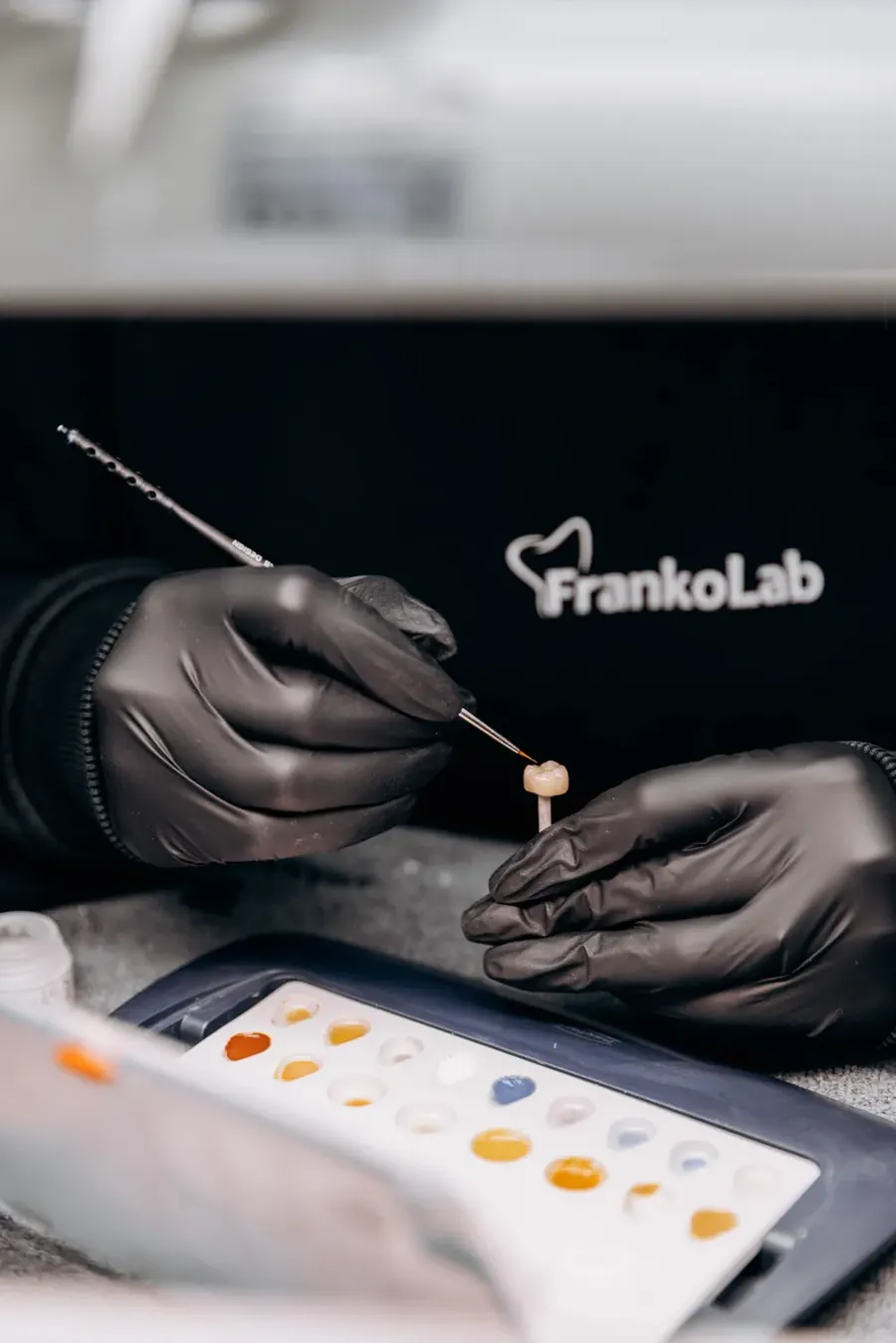
A FrankoLab dental technician works on a crown in the clinic’s in-house laboratory
01

Treatment under a surgical microscope

We carry out all procedures involving perforations under a high-powered dental microscope (Carl Zeiss / Leica / Scanner). A magnification of 20–30 times enables us to precisely locate the defect and view the perforation site in detail—beyond what is visible to the naked eye—thereby sealing it hermetically and minimising the risk of complications.

02

Accurate on-site diagnosis

Our in-house J. Morita 3D Accuitomo 170 CT scanner enables us to assess the size, location of the perforation and the condition of the surrounding bone tissue in a single visit. This enables us to draw up the most accurate treatment plan possible and select the optimal method for closing the perforation in the tooth root.

03

Modern biocompatible materials

We use tried-and-tested materials (MTA, bioceramics) which ensure a reliable seal, promote bone regeneration and have a high success rate in the long term.

04

A comprehensive approach to tooth preservation

If a perforation is combined with other endodontic issues — such as root canal retreatment, resorption or the need for surgical access — we address all these issues within a single framework, managed by a single team of dentists.

05

Predicted outcome and tooth preservation

Our aim is not simply to fill a cavity, but to preserve the tooth for many years to come. Thanks to the use of a microscope, modern protocols and our experience, we achieve a high success rate, even in complex (particularly repeat) cases.

Frequently asked questions about closing a root perforation

A root perforation does not always produce obvious symptoms straight away. The most common signs include pain when biting down or a constant dull ache in the area of the tooth, swelling or tenderness of the gums adjacent to the affected tooth, a gum abscess with pus discharge, and discolouration of the tooth. Sometimes a root perforation is only detected on an X-ray — during a routine check-up or root canal treatment. If you experience even one of these symptoms, see your dentist without waiting for the pain to get worse.
In most cases, recovery is possible if treatment is sought in good time. The prognosis depends on the location of the defect, its size, how long it has been present, and the condition of the surrounding bone tissue. Perforations in the coronal and middle thirds of the root, diagnosed at an early stage, respond well to treatment. Neglected cases involving extensive bone destruction are more complex, but even in such cases we try to find a solution before recommending extraction.
The procedure will be carried out under local or general anaesthesia — the patient will feel no pain during treatment. Once the anaesthetic has worn off, you may experience slight discomfort in the treated area, which can be easily relieved with standard painkillers and will subside within a few days.
The duration depends on the complexity of the case. A simple repair of a fresh perforation in a single visit takes 60–90 minutes. More complex cases — involving active inflammation, the need for a temporary filling or surgical access — require 2–3 visits.
A microscope is not merely desirable; it significantly improves the accuracy and predictability of the outcome. A defect invisible to the naked eye can be assessed, cleaned and sealed with maximum airtightness under a microscope. Treating a perforation without a microscope is like working blind, which increases the risk of incomplete closure and recurrence.
MTA (Mineral Trioxide Aggregate) — a material based on mineral trioxide aggregate, which is the gold standard in the treatment of root perforations. Its key properties include: high biocompatibility, the ability to set in a moist environment (which is critical for root canal conditions), stimulation of surrounding tissue regeneration, and reliable, long-lasting sealing. It is precisely these characteristics that make MTA the material of choice for treating root perforations.
Perforation is most often a complication of endodontic treatment or the result of advanced tooth decay. Prevention involves regular check-ups every six months, prompt treatment of tooth decay without delay, and choosing an endodontist with extensive clinical experience and modern equipment. Root canal treatment under a microscope with CT guidance significantly reduces the risk of iatrogenic complications from the outset.

Book a consultation

Book a consultation and one of our managers will contact you shortly

+380 50 011 20 00

FrankoLab Clinic dental surgery

A healthy smile starts with the right decision

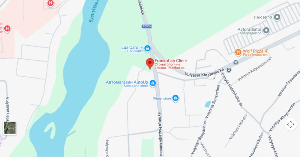
  • 77460, Ivano-Frankivsk, Cherniiv village, 2B Nadvirnianska Street
  • Mon–Sat 9.30 am–7.00 pm, Sun 10.00 am–7.00 pm
  • +380 50 011 20 00
Book a consultation
Map: 77460, Ivano-Frankivsk, village of Cherniiv, 2B Nadvirnianska Street