Deep caries affecting the pulp
When tooth decay reaches the pulp chamber, the nerve becomes inflamed, causing severe or throbbing pain. Root canal treatment is required.

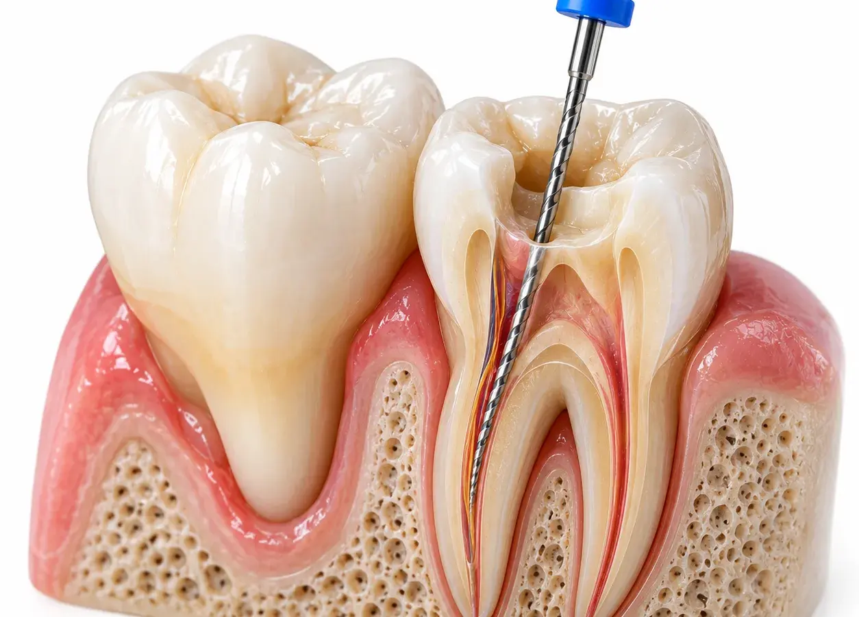
Premolars, or small molars (‘fours’ and ‘fives’), are situated between the canines and molars. They are important for chewing food and for forming and maintaining the bite. Despite being smaller than molars, they are just as prone to tooth decay, pulpitis and other conditions requiring endodontic treatment.
Endodontic treatment of a premolar involves treating the internal tissues of the tooth: removal of inflamed or infected pulp, cleaning and disinfection of the root canals, and sealing them with biocompatible materials. The procedure makes it possible to preserve the natural tooth, even in complex clinical cases. At FrankoLab Clinic, we carry out endodontic treatment of premolars using a Carl Zeiss / Leica / Scanner dental microscope and digital diagnostics without involving external clinics — to ensure maximum precision and effective results.
When tooth decay reaches the pulp chamber, the nerve becomes inflamed, causing severe or throbbing pain. Root canal treatment is required.
Inflammation of the premolar pulp is accompanied by sensitivity to hot and/or cold and night-time pain. Endodontic treatment eliminates the source of inflammation and preserves the tooth.
If the infection has spread beyond the root into the surrounding tissues, pain may occur when biting down, along with swelling or a fistula on the gums. Root canal treatment of a premolar helps to eliminate the source of the infection.
A blow, a chip or a deep crack can damage the pulp chamber and lead to acute inflammation. Timely endodontic treatment of a premolar saves the tooth.
Discolouration is a sign of pulp necrosis, which requires an examination of the root canals and, if necessary, treatment of the premolar.
Such pathological conditions are chronic inflammatory processes that are treated endodontically (sometimes in combination with microsurgical intervention).
Before fitting a crown onto a severely damaged premolar, root canal treatment and high-quality filling of the canals are usually required.
The cost of endodontic treatment for a premolar at FrankoLab Clinic in Ivano-Frankivsk includes a comprehensive range of services: diagnosis, anaesthesia, root canal preparation and filling, and restoration of the tooth crown. The final price is determined on a case-by-case basis following an examination and X-ray diagnosis. It depends on the number of canals in the specific tooth, the complexity of the case and the condition of the tissues. The dentist will quote the exact amount before treatment begins, with no hidden charges. The cost of treatment can be split into manageable instalments without any extra charges — please check with the administrator for the current payment terms for premolar treatment.
The dentist will quote the final cost following an examination and diagnosis. Payment in instalments is available. Full price list for the clinic
An endodontist examines the tooth, assesses the symptoms and the severity of the pain. We take a targeted X-ray or perform a low-dose CT scan using the J. Morita 3D Accuitomo 170 to accurately determine the anatomy of the premolar root canals. Based on the results, we make a diagnosis and draw up a treatment plan.
For premolar root canal treatment, we administer a modern local anaesthetic. After checking the level of sensitivity, we fit a cofferdam: a latex or latex-free isolation system that protects the tooth from saliva and ensures the surgical site is completely sterile.
Under the guidance of a dental microscope, the dentist gains access to the pulp chamber and removes the affected pulp along with the neurovascular bundle. The microscope makes it possible to detect even additional and very fine canals, which cannot be seen with the naked eye during endodontic treatment of premolars.
We enlarge and mechanically clean the root canals using nickel-titanium instruments. At the same time, we carry out thorough rinsing with antiseptic solutions to ensure complete chemical disinfection. We monitor the length of each root canal using an apex locator.
During endodontic treatment of a premolar, once the root canals have been thoroughly cleaned and shaped, we fill them tightly with gutta-percha and sealer. A hermetically sealed filling prevents bacteria from re-entering and ensures a long-lasting result.
To complete the root canal treatment of a premolar, we place a temporary or permanent filling to restore the tooth’s shape and function. The dentist provides care advice and arranges a follow-up X-ray. In cases of significant damage, we recommend having a crown fitted to protect the tooth.
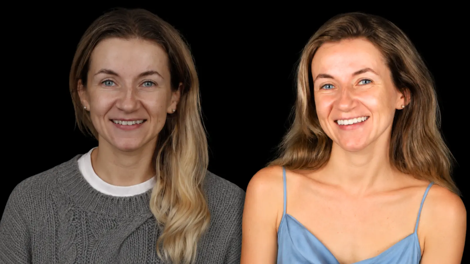
The patient presented with complaints of discolouration of her front teeth, numerous old composite restorations and an unsatisfactory smile. During the clinical examination, discolouration of the teeth was identified, along with restorations that had lost their functionality and aesthetic appearance.
Following digital planning, a decision was made to carry out aesthetic restoration of the upper incisors using refractive veneers.
The restorations
are made entirely from ceramic materials, without any metal framework, which is one of the most aesthetically pleasing techniques in modern dentistry. This method allows for the most accurate reproduction of the natural translucency, colour depth and optical properties of natural enamel, ensuring a flawless result.
Discolouration has been removed and old, unaesthetic restorations have been replaced. The natural shape, proportions and harmony of the front teeth have been restored. Thanks to refractive veneers, the patient achieved a natural, bright and aesthetically pleasing smile that closely mimics natural teeth, combining durability, functionality and premium aesthetics.

Diana Kruk
08.07.2026
The clinic and service are of the highest standard – keep up the good work!
Ksenia Savenko
23.06.2026
I am very grateful to the whole FrankoLab team for the experience I’ve had with you. You are incredible! I wish you continued growth and prosperity, because the smiles you create are not only beautiful but also genuine, as seen on the faces of your grateful clients.
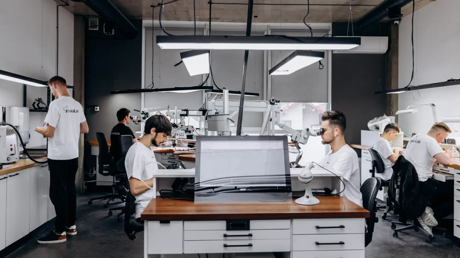
Premolars often have a complex root canal anatomy (particularly the upper ones — two or more canals, bends, and additional branches). Under a Carl Zeiss / Leica / Scanner microscope with 20–30x magnification, the dentist locates all the root canals, cleans them thoroughly and seals them hermetically, minimising the risk of residual infection.
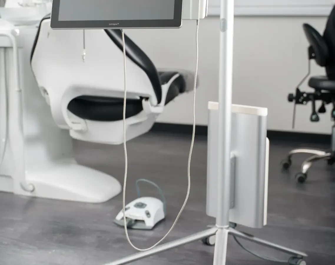
The J. Morita 3D Accuitomo 170 CT scanner enables us to accurately assess the number and configuration of the premolar root canals, the thickness of the root walls and the presence of any complications even before treatment begins. This makes the treatment predictable.
The endodontists at our clinic regularly carry out both initial treatment and retreatment of premolars, the removal of instrument fragments and the repair of perforations. Many teeth that other dental practices have recommended for extraction can be preserved for a long time to come.
We use the latest international protocols for root canal treatment and obturation using biocompatible materials. This ensures long-lasting results and reduces the risk of re-infection.
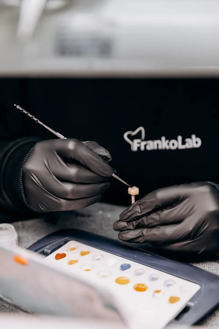
Following endodontic treatment of a premolar, the tooth can be restored immediately with a high-quality filling or a crown at our clinic. FrankoLab’s in-house dental laboratory guarantees speed and quality control at every stage.
Book a consultation and one of our managers will contact you shortly
Book a consultation and one of our managers will contact you shortly
Would you like a remote consultation? Online consultation with a dentist
Share your experience of your treatment — your review will appear on the website once it has been verified by an administrator.