EN
Apical microsurgery

Why choose FrankoLab:

  1. 01 Removal of cysts and granulomas without tooth loss
  2. 02 Microsurgical precision under an operating microscope
  3. 03 We treat cases where root canal retreatment is ineffective

Apical microsurgery in Ivano-Frankivsk

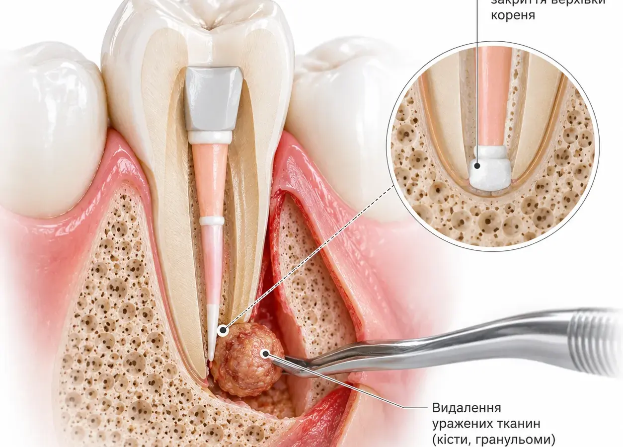
Apical microsurgery is a tooth-preserving microsurgical procedure, during which the dentist gains direct access to the apex of the tooth root via a small incision in the gums, removes the affected tissue (such as a cyst or granuloma) and hermetically seals the root apex. Unlike root canal retreatment, apical surgery acts directly on the site of inflammation, bypassing the limitations associated with the anatomy of the root canal. It is prescribed when conservative endodontic treatment is ineffective or impossible. At FrankoLab Clinic, we perform root-apex resection under a Carl Zeiss / Leica M655 / Scanner surgical microscope, which ensures precision, minimal trauma and a predictable outcome.

Apical microsurgery is recommended when root canal treatment (initial or retreatment) has proved ineffective or is contraindicated, and the tooth is of sufficient strategic value to warrant preservation.

The decision on whether treatment is possible is always made by an endodontist or oral surgeon following a clinical examination, targeted X-rays and a CT scan.

Indications for apical microsurgery

Persistent or recurrent apical periodontitis

Apical microsurgery makes it possible to eliminate the source of inflammation at the tip of the root when high-quality treatment or retreatment of the root canals has not been successful, and symptoms or radiographic signs of inflammation persist.

Inability to gain orthodral access to the root canals

Root canal treatment is helpful in cases of complex anatomy (severely curved canals, obliteration, additional canals), where there are posts, inlays or broken instruments that cannot be safely removed, or where there is a risk of damaging the tooth if retreatment is attempted via the usual access through the canals.

Presence of a cyst or granuloma at the apex of the root

Microsurgical intervention allows the pathological lesion to be completely removed along with the affected part of the root and the canal to be hermetically sealed, ensuring bone tissue healing and tooth preservation.

Extruded filling material for the root apex

When material that has extended beyond the apex causes chronic inflammation and symptoms, apical microsurgery makes it possible to remove this material and the affected tissue, followed by retrograde filling.

Perforation of the root or the floor of the pulp chamber

If it is not possible to seal a perforation effectively from within the root canal, microsurgical access allows the defect to be repaired, the inflamed tissue to be removed and the root canal to be hermetically sealed from the apical end.

Teeth with crowns, bridgework or prosthetic restorations

Apical microsurgery makes it possible to save a tooth and avoid removing a high-quality prosthetic restoration when root canal retreatment through the crown is technically difficult or undesirable.

Apical root fracture

In the case of an isolated fracture of the root apex, resection of the damaged portion followed by retrograde filling makes it possible to preserve the tooth and eliminate the source of infection.

How much does apical microsurgery cost?

The cost of apical microsurgery at FrankoLab Clinic in Ivano-Frankivsk depends on the complexity of the clinical case, the size of the pathological lesion, the need for bone grafting and the scope of associated procedures. We provide a transparent quote following a consultation and a CT scan — with no hidden surcharges during the process. If required, payment plans are available with no additional charges.

The dentist will quote the final cost following an examination and diagnosis. Payment in instalments is available. Full price list for the clinic

Stages of apical microsurgery on a tooth

01

Preparation for apical microsurgery

We carry out CT scans using the J. Morita 3D Accuitomo 170 to accurately assess the location and size of the lesion, the condition of the bone tissue and the root anatomy. We assess your general state of health and, if necessary, prescribe preparatory medication. We draw up a treatment plan.

02

Anaesthesia and access

We offer a choice of local or general anaesthesia. We make a small incision in the gums and lift the muco-osseous flap to gain direct access to the root apex. The entire subsequent procedure is carried out under a surgical microscope.

03

Root apex resection

We remove the affected bone tissue and the pathological focus. We resect the tip of the tooth root (performing an apicoectomy — removal of the tip of the tooth root) — usually 2–3 mm — along with the unfilled or infected portion.

04

Retrograde preparation and filling

We prepare a cavity at the root tip, thoroughly rinse it with antiseptics and hermetically seal the apex with a biocompatible material — usually MTA, bioceramics or other modern alternatives. The technique of apical microsurgery prevents re-infection from the root canal.

05

Suturing and finishing

We irrigate the surgical site and, if necessary, place a bone substitute into the defect area to accelerate regeneration. We reposition the flap and apply sutures. The operation takes approximately 60–90 minutes.

06

Post-operative period

We take a follow-up X-ray and provide recommendations (including, amongst other things, antibiotics if indicated, painkillers and antiseptics). We remove the stitches after 5–7 days. We usually schedule follow-up examinations and X-rays to assess healing at 3, 6 and 12 months.

Our patients’ results

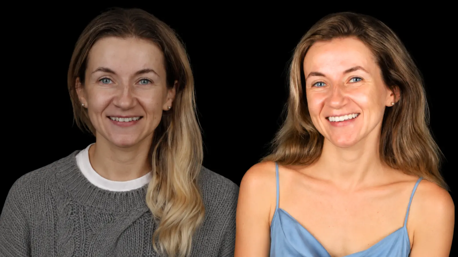
Case Study 3: Composite Restorations

Diagnosis:

The patient presented with complaints of discolouration of her front teeth, numerous old composite restorations and an unsatisfactory smile. During the clinical examination, discolouration of the teeth was identified, along with restorations that had lost their functionality and aesthetic appearance.

Treatments:

Following digital planning, a decision was made to carry out aesthetic restoration of the upper incisors using refractive veneers.
The restorations
are made entirely from ceramic materials, without any metal framework, which is one of the most aesthetically pleasing techniques in modern dentistry. This method allows for the most accurate reproduction of the natural translucency, colour depth and optical properties of natural enamel, ensuring a flawless result.

Result:

Discolouration has been removed and old, unaesthetic restorations have been replaced. The natural shape, proportions and harmony of the front teeth have been restored. Thanks to refractive veneers, the patient achieved a natural, bright and aesthetically pleasing smile that closely mimics natural teeth, combining durability, functionality and premium aesthetics.

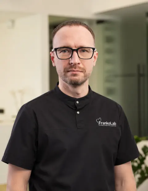
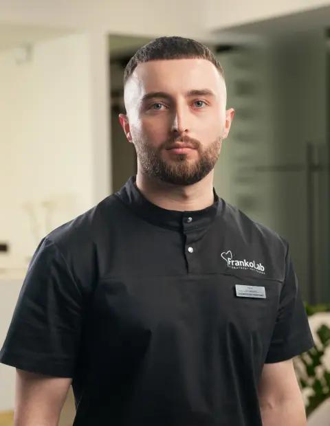
Doctors providing treatment

Patient reviews

4,9 Google rating All reviews on Google

Diana Kruk

08.07.2026

The clinic and service are of the highest standard – keep up the good work!

Ksenia Savenko

23.06.2026

I am very grateful to the whole FrankoLab team for the experience I’ve had with you. You are incredible! I wish you continued growth and prosperity, because the smiles you create are not only beautiful but also genuine, as seen on the faces of your grateful clients.

FrankoLab Dental Laboratory

Not sure if your tooth can be saved? Book a consultation at FrankoLab Clinic — we’ll carry out a CT scan and determine whether apical microsurgery is recommended for you.

Why do people choose FrankoLab Clinic for apical microsurgery?

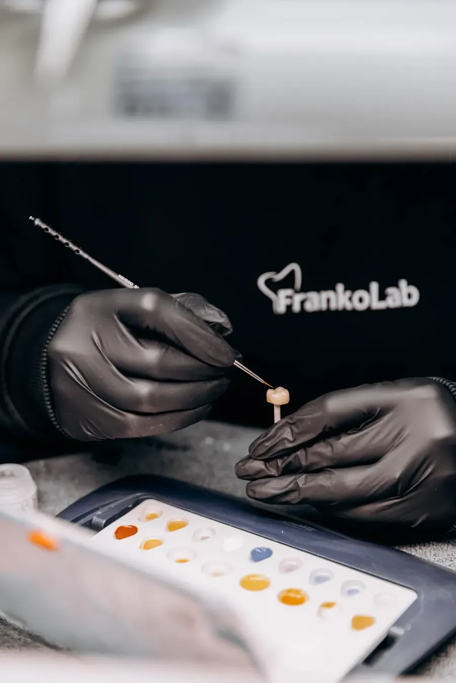
A FrankoLab dental technician works on a crown in the clinic’s in-house laboratory
01

Surgery performed exclusively under a microscope

We use Carl Zeiss, Leica M655 and Scanner surgical microscopes. A microscope is not an option, but the standard: it allows us to see the root apex and the surgical field in detail, beyond the capabilities of the naked eye, and to work with a precision that determines the quality of the result.

02

Precise CT diagnostics prior to surgery

A CT scan using the J. Morita 3D Accuitomo 170 allows us to determine the exact location of the lesion, its size and position relative to neighbouring structures even before the operation — and to plan the procedure without any surprises.

03

Experience in complex and repeat cases

We undertake surgical procedures following unsuccessful previous attempts, in cases involving retained instrument fragments, perforations and large cysts. If other dental practices have only offered extraction, please contact us.

04

A complete course of treatment under one roof

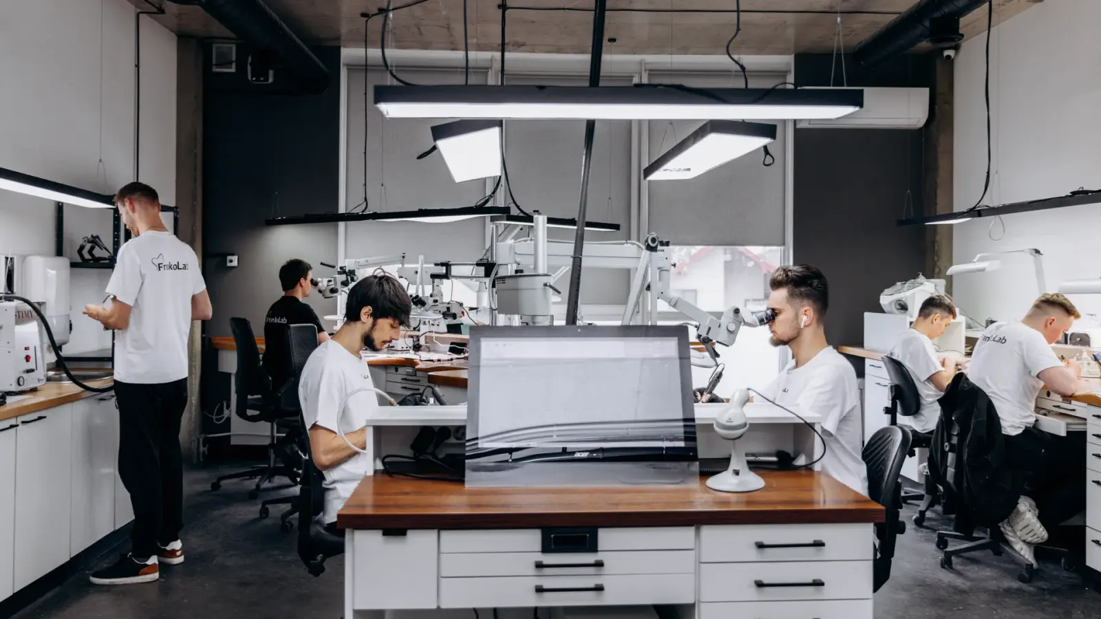
At FrankoLab Clinic, you can go through the entire process — from diagnosis and planning to apical microsurgery, monitoring of healing and subsequent tooth restoration — without needing to visit other clinics. Everything takes place within a single system, featuring our own dental laboratory and a team of specialists.

05

Transparent pricing and dental tourism

You will receive a detailed quote before treatment begins. We welcome patients from all over Ukraine and abroad: a free transfer service, online consultations and thorough planning help to minimise the number of visits.

Frequently asked questions about apical microsurgery

Root canal treatment involves working inside the tooth: the dentist treats and fills the canal via the crown. Apicoectomy is performed externally: through an incision in the gums, the dentist gains direct access to the root tip and surgically removes the pathological focus. These two approaches do not compete — they complement one another. Microsurgery is prescribed when conservative treatment options have been exhausted or are not feasible.
On the day before your procedure, we recommend that you eat a good meal and get a good night’s sleep, and avoid alcohol for at least 24 hours. If you are taking anticoagulants or any other medication, you must inform your dentist. You should brush your teeth immediately before the operation. Detailed recommendations on how to prepare for apical microsurgery will be provided by the doctor during your consultation, taking your condition into account.
We carry out the procedure under anaesthesia (local or general) — the patient feels no pain during the procedure. Once the anaesthetic has worn off, you may experience mild discomfort, swelling and tenderness in the area of the operation — this is a normal reaction of the tissues. These sensations can be effectively managed with the prescribed painkillers and will subside within a few days.
Usually 60–90 minutes, depending on the complexity of the case, the location of the tooth and the size of the affected area. Surgery on molars or involving large cysts sometimes takes longer.
Stitches are usually removed 7–10 days after the operation. External healing of the gums is complete within 2–4 weeks. Full regeneration of bone tissue at the site of the extracted tooth takes from several months to a year — this is monitored via X-rays during follow-up appointments.
Most patients return to their normal routine as early as the second or third day. On the first day, vigorous mouth rinsing is not permitted. You must strictly follow the dentist’s recommendations regarding the recovery period. Swelling and bruising around the surgical site are normal and will subside within 3–5 days.
When performed correctly, complications are rare. Possible complications include: temporary numbness or increased sensitivity in the area of the operation, slight separation of the sutures, and, rarely, wound infection. Serious complications (damage to adjacent teeth or nerves) are minimised through precise CT diagnostics and working under a microscope.
The root apex is a microscopic structure. Without magnification, it is impossible to see fine details: additional canals, cracks, or the quality of the retrograde filling. A microscope transforms the procedure from an ‘approximate’ one into a precise and predictable one. This is precisely why apicoectomy without a microscope is considered, by modern standards, an outdated and ineffective approach.
Yes. Absolute contraindications: severe systemic diseases in the decompensated stage, blood clotting disorders, a recent heart attack or stroke, and cancer of the maxillofacial region. Relative contraindications: active inflammation in the acute stage, poor oral hygiene, smoking (increases the risk of healing complications), certain chronic conditions — these require prior treatment. Please enquire about the full list during your consultation.

Book a consultation

Book a consultation and one of our managers will contact you shortly

+380 50 011 20 00

FrankoLab Clinic dental surgery

A healthy smile starts with the right decision

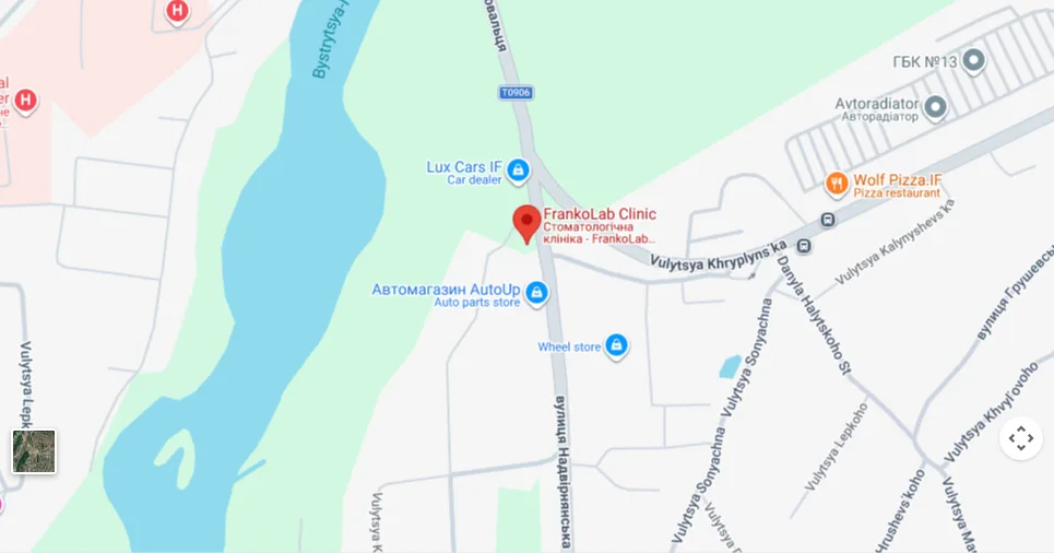
  • 77460, Ivano-Frankivsk, Cherniiv village, 2B Nadvirnianska Street
  • Mon–Sat 9.30 am–7.00 pm, Sun 10.00 am–7.00 pm
  • +380 50 011 20 00
Book a consultation
Map: 77460, Ivano-Frankivsk, village of Cherniiv, 2B Nadvirnianska Street