EN
Segmental CBCT scan (3–4 teeth)

Why choose FrankoLab:

  1. 01 Detailed 3D analysis of the selected section of the dentition
  2. 02 Required for dental implants and endodontic treatment
  3. 03 High precision with minimal radiation exposure

Segmental CT scan (3–4 teeth) in Ivano-Frankivsk

Traditional X-rays often do not provide a complete picture of the condition of the teeth, their root systems and the surrounding tissues. Without a detailed three-dimensional analysis of the structure of the teeth, it is impossible to plan implant surgery, endodontic treatment, orthodontic procedures or complex surgical interventions.

Segmental CT scan – 3D diagnosis of a local area comprising 3–4 teeth.

This is a high-tech diagnostic procedure that produces extremely detailed three-dimensional images. The scanning area during a CT scan is. The procedure is carried out using a modern cone-beam computed tomography scanner. Specialised software generates an accurate, detailed 3D model of the area under examination. This enables the dentist to detect even the smallest hidden abnormalities, cracks or inflammatory processes.

Advantages of segmental CT

Ultra-high diagnostic accuracy.

Segmental CT provides highly detailed 3D images. This makes it possible to detect microscopic abnormalities, the finest cracks in the enamel, hidden root canals and periapical changes that cannot be seen on two-dimensional images.

Maximum safety for your health.

The state-of-the-art equipment uses the lowest possible radiation dose, which is 8–10 times lower than that of a spiral CT scan of the jaws. The procedure is safe for patients, including children, and can be carried out several times a year where medically indicated.

Lightning-fast scanning speed.

The procedure takes a record-breaking short time – just 10–15 seconds – making it as comfortable as possible even for patients with claustrophobia or children. The ultra-fast scan completely minimises the risk of artefacts caused by the patient’s involuntary movements.

Complete comfort throughout the procedure.

The patient assumes a comfortable, natural position in the spacious, ergonomically designed CT scanner chair. The entire scanning process is painless, silent and causes no discomfort whatsoever. No special preparation is required.

Affordable diagnostic costs.

A CT scan costs less than a full jaw tomography, whilst providing absolutely all the clinical information required for an accurate diagnosis and successful treatment of a specific problem area. The competitive price makes diagnosis affordable.

High-quality treatment planning.

High-quality 3D images enable the dentist to plan procedures with precision: implant placement, determining the optimal size and position of implants; extraction of impacted wisdom teeth; and endodontic treatment.

Clinical indications for segmental CT scans

Detailed planning for dental implants.

Segmental CT is the gold standard and an essential procedure for high-quality implant planning. The procedure allows the structures of the teeth, bone tissue and jaws to be assessed with micron-level precision.

Detection of hidden and secondary caries.

Traditional two-dimensional X-rays are often unable to detect carious lesions located between teeth or in hard-to-reach areas. A CT scan of 3–4 teeth allows even the early stages of hidden caries to be diagnosed.

Complex endodontic root canal treatment.

During root canal treatment, particularly in complex clinical cases, CT dental diagnostics provides information to ensure the procedure is effective and to minimise the risk of recurrent infection.

Comprehensive diagnosis following maxillofacial injuries.

In the event of traumatic injuries to the dentally-maxillary system, it is extremely important to assess the nature and extent of the damage in detail. Dentistry requires the most accurate diagnosis in order to select the correct treatment approach.

Assessment of complex cases prior to wisdom tooth extraction.

Accurate dental diagnosis using CT scans helps to determine in detail the spatial position of impacted and malpositioned wisdom teeth, as well as the thickness of the bone tissue, for the purpose of planning the surgical extraction procedure.

Affordable pricing for professional segmental CT scans

Segmental CT scans at FrankoLab Clinic combine the highest quality of diagnosis with fair pricing, with no hidden surcharges or commissions. Our clinic carries out dental CT scans in Ivano-Frankivsk, with transparent pricing for procedures. The cost of dental diagnostics includes a consultation with an experienced radiologist, the scan itself, professional analysis of the results and a detailed report with recommendations.

The dentist will quote the final cost following an examination and diagnosis. Payment in instalments is available. Full price list for the clinic

A detailed guide to the segmental CT scan procedure

Cone-beam CT scans at FrankoLab Clinic are performed using state-of-the-art equipment that meets strict international standards. Our specialists, with many years of experience, ensure the highest quality of diagnosis and comfort for every patient. Results are provided on the day of your appointment, allowing treatment to begin without unnecessary delays.

01

Initial consultation and treatment planning.

Our attentive dentist carries out a thorough initial examination of the oral cavity, takes a detailed medical history of the patient’s condition, and analyses their complaints and symptoms to determine whether a segmental CT scan is required to diagnose a specific dental problem.

02

Preparatory stage prior to scanning.

The patient removes all metal objects that may cause artefacts in the image. The nurse explains the rules to follow during the scan, the importance of remaining still, and helps the patient to get into a comfortable position. A dental CT scan does not require any special preparation.

03

Precise positioning in the CT scan.

The patient assumes a comfortable position in a specially designed chair attached to a modern CT scanner, after which their head is gently secured with soft headrests and stabilising devices to ensure complete immobility during the scan. The technical parameters of the scan are configured.

04

On-site scanning.

The scan takes just 5–10 seconds, during which the patient must remain completely still and breathe calmly and naturally. The tomograph rotates around the patient’s head, producing a series of high-quality X-ray images from various angles. The procedure is fully automated.

05

Professional processing and analysis of results.

The data obtained is immediately processed using state-of-the-art specialised software, which creates an extremely detailed, high-quality three-dimensional model of the anatomical area under examination. A radiologist analyses the resulting 3D reconstructions and identifies any abnormalities.

06

Receiving results and consultation.

The patient receives the results of the examination (digital 3D images, a printed professional report from a radiologist with a detailed description of the findings and clinical recommendations). The dentist explains all aspects of the examination results in detail and draws up a treatment plan.

Japanese CT equipment ensures accurate dental diagnosis.

FrankoLab Clinic uses the state-of-the-art Japanese Morita Accuitomo 170 CT scanner, which provides maximum image detail with high resolution whilst minimising radiation exposure to the patient. Dental CT scan results are provided in a modern digital format, allowing for detailed viewing in specialised software, so that they can be saved permanently.

Doctors providing treatment

Patient reviews

4,9 Google rating All reviews on Google

Diana Kruk

08.07.2026

The clinic and service are of the highest standard – keep up the good work!

Ksenia Savenko

23.06.2026

I am very grateful to the whole FrankoLab team for the experience I’ve had with you. You are incredible! I wish you continued growth and prosperity, because the smiles you create are not only beautiful but also genuine, as seen on the faces of your grateful clients.

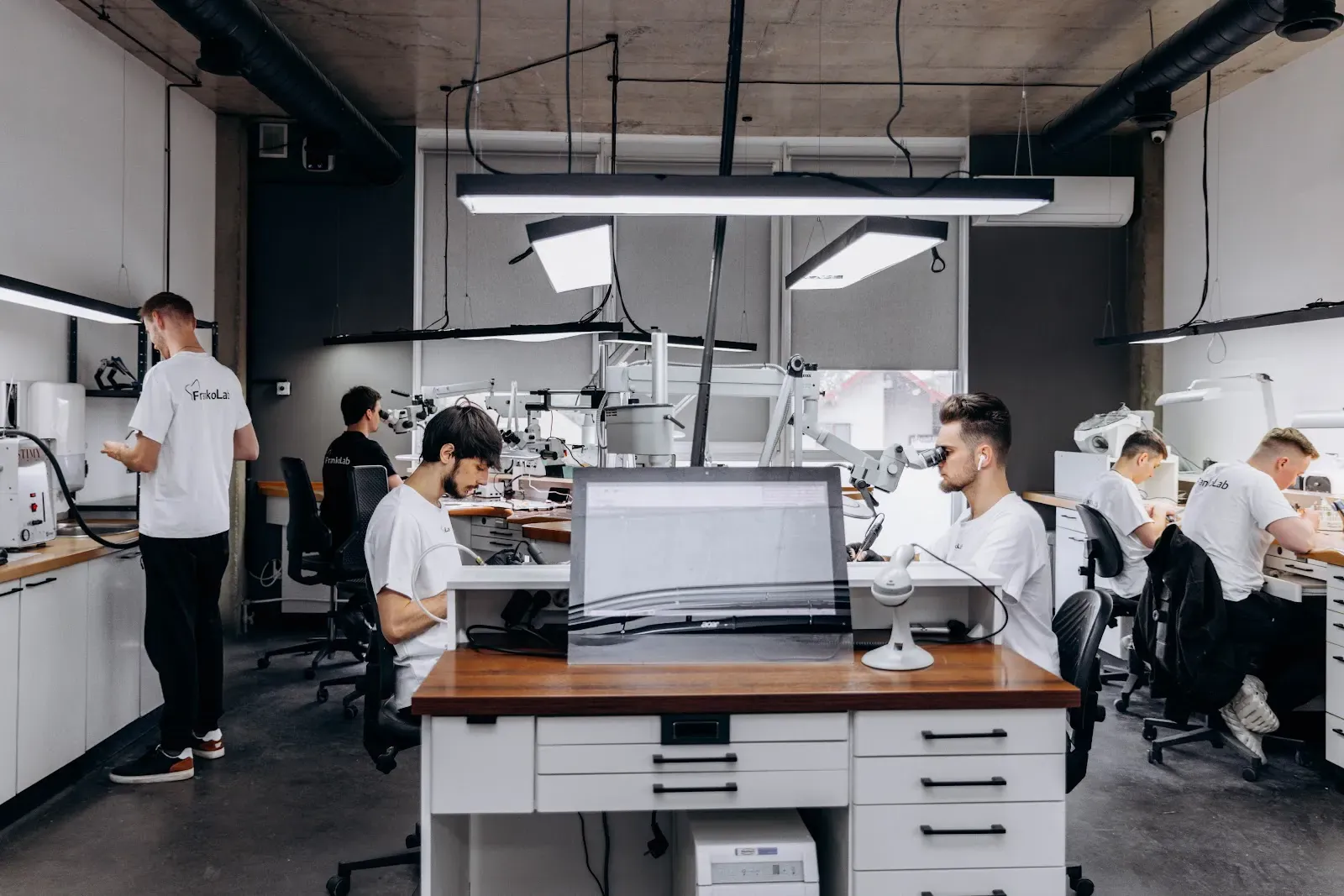
In-house laboratory

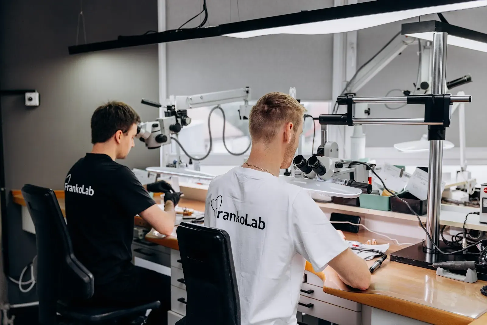
FrankoLab’s main advantage is its own in-house dental laboratory.

As all our production facilities are located on-site, we can fully control the manufacturing process for restorations of any complexity, develop solutions quickly and guarantee long-term results.

In our digital dental laboratory, metal-free prosthetic restorations are manufactured using leading CAD/CAM technologies and the highest-quality European materials.

FrankoLab Dental Laboratory

Why do patients choose FrankoLab Clinic for segmental CT scans?

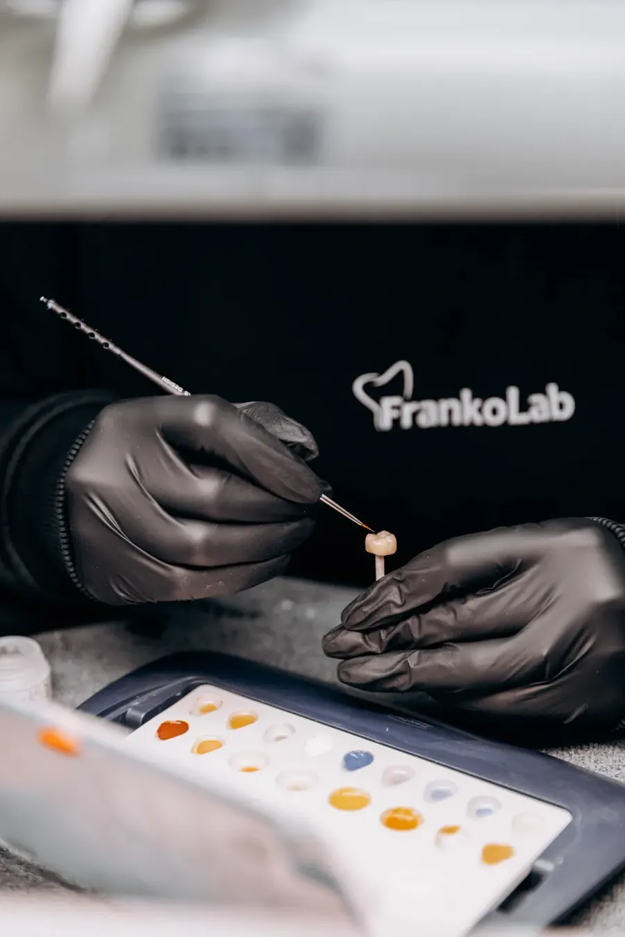
A FrankoLab dental technician works on a crown in the clinic’s in-house laboratory
01

We use state-of-the-art, world-class equipment.

FrankoLab Clinic is equipped with a latest-generation CT scanner, which ensures high-precision segmental CT scans whilst minimising the patient’s radiation exposure. Dental diagnostics are carried out using state-of-the-art technology.

02

We have a team of experienced dentists working here.

The staff at our dental clinic have many years’ practical experience in managing complex clinical cases. Accurate diagnosis is ensured by in-depth knowledge and expert use of specialist software.

03

Maximum patient comfort and speedy service.

Dental CT scans are carried out without queues or waiting times, by appointment at a time convenient for the patient. Results are prepared quickly once the scan is complete, allowing treatment planning and treatment to begin immediately.

04

Transparent pricing and a guarantee of a high-quality examination.

Our clinic carries out diagnostic procedures at a fair price. We provide detailed consultations on the results. Diagnostics include a full range of services at a fixed price with no hidden surcharges or additional costs.

Frequently asked questions about segmental CT scans

A segmental CT scan is a computed tomography scan that produces a three-dimensional image of 3–4 teeth within the scan area. Dental diagnostics allow for a detailed assessment of the condition of the root system and bone tissue, and the detection of hidden pathologies. 3D images of the teeth are used to plan implant placement, endodontic treatment and surgical procedures.
Dental CT scans are completely safe for your health, as they use a minimal dose of radiation. The safety of CT scans has been confirmed by international studies; the procedure can be carried out several times without causing any harm to the body. Diagnostics are carried out in compliance with radiation safety regulations in accordance with approved standards.
A local CT scan takes just 10–15 seconds. The total time spent at the clinic is 15–20 minutes, including preparation and positioning. Dental diagnostics are carried out quickly and comfortably, without any pain. The results are processed within an hour, and the patient receives a report and digital images on the day of their appointment.
A CT scan for implant placement provides information on the volume and density of bone tissue, as well as the position of nerves and blood vessels, which is crucial for the successful placement of implants. A 3D image allows us to determine the size and type of implant, as well as the optimal angle for its insertion. Dental diagnostics minimises the risk of complications and ensures a predictable outcome for the implant procedure.
A segmental CT scan provides three-dimensional visualisation, unlike conventional X-rays, which show a two-dimensional image. Compared to a full CT scan of the jaws, a localised scan focuses on a specific area, reducing both the radiation dose and the cost of the procedure. This diagnostic method reveals details that are not accessible to other examination methods.

Further details: segmental CBCT scan (3–4 teeth)

Computed tomography (CT) of the teeth, also known as a 3D scan or CT scan, is one of the main diagnostic methods in modern dentistry. It enables the dentist to analyse both individual teeth and their tissues, as well as the overall condition of the jaw, and to detect lesions and inflammation from various angles.

Computed tomography (CT) scans of the jaw are used in many areas of dentistry. At FrankoLab Clinic, we take a comprehensive approach to treatment, where each specialist – such as a general dentist, orthodontist, periodontist and implant surgeon – may be involved in the treatment of a single patient. A CT scan is the most informative method for each of them.

With the help of a CT scan, it is possible to:

  • Assess the condition of the root canals.
  • Diagnosing cysts and granulomas.
  • Detect inflammation.
  • Detecting tooth buds in children and adolescents.
  • Book an extraction or other surgical procedures.
  • Diagnose hidden tooth decay.
  • Monitor the progress of previous treatment.
  • Quick diagnosis of jaw injuries.

A dental CT scan is a quick, safe and non-invasive procedure for the patient. They simply need to remove any metal jewellery before the procedure, which can be done directly in the X-ray room. The FrankoLab clinic is equipped with the most modern and safest expert-level CT scanner, the MORITA ACCUITOMO F170, which quickly produces clear images so that the patient and the dentist can view the results of the examination within a few minutes.

We have a unified system for storing and viewing CT scans. Each dentist can review the scan results together with the patient. In addition, patients can receive their CT scans in digital format on a disc or via email.

Book a consultation

Book a consultation and one of our managers will contact you shortly

+380 50 011 20 00

FrankoLab Clinic dental surgery

A healthy smile starts with the right decision

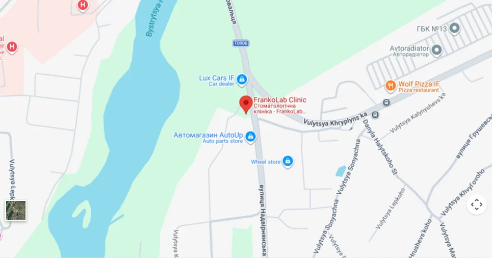
  • 77460, Ivano-Frankivsk, Cherniiv village, 2B Nadvirnianska Street
  • Mon–Sat 9.30 am–7.00 pm, Sun 10.00 am–7.00 pm
  • +380 50 011 20 00
Book a consultation
Map: 77460, Ivano-Frankivsk, village of Cherniiv, 2B Nadvirnianska Street